Search This Blog

This is default featured slide 1 title

Go to Blogger edit html and find these sentences.Now replace these sentences with your own descriptions.

This is default featured slide 2 title

Go to Blogger edit html and find these sentences.Now replace these sentences with your own descriptions.

This is default featured slide 3 title

Go to Blogger edit html and find these sentences.Now replace these sentences with your own descriptions.

This is default featured slide 4 title

Go to Blogger edit html and find these sentences.Now replace these sentences with your own descriptions.

This is default featured slide 5 title

Go to Blogger edit html and find these sentences.Now replace these sentences with your own descriptions.

Monday, February 28, 2011

Histology and Explanation of chondroma

Histology and Explanation of chondroma

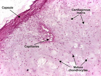
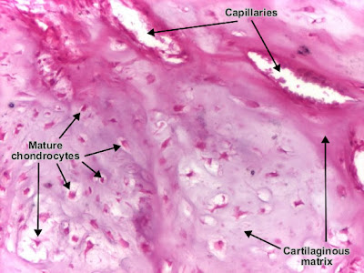
Chondroma is a benign cartilaginous tumor, encapsulated, with a lobular growing pattern. Tumor cells (chondrocytes, cartilaginous cells) resemble normal cells and produce the cartilaginous matrix (amorphous, basophilic material). Characteristic are the vascular axes within the tumor, which make the distinction with normal hyaline cartilage.

Histology and Explanation of chondroma

Top 10 Medical Quotations of the Week


"Nothing is more fatal to health than over care of it."
~ Ben Franklin

"Unless the doctor of today becomes the dietitian of tomorrow, the dietitian of today will become the doctor of tomorrow."
~ Dr. Alexis Carrol

"I learned a long time ago that minor surgery is when they do the operation on someone else, not you."
~ Bill Walton

"Treat the patient, not the X-ray."
~ James M. Hunter

"He's the best physician that knows the worthlessness of the most medicines."
~ Benjamin Franklin

"Finish last in your league and they call you idiot. Finish last in medical school and they call you doctor."
~ Abe Lemons

"The desire to take medicine is perhaps the greatest feature which distinguishes man from animals."
~ William Osler

"Classifying thoughts, feelings and behaviors as diseases is a logical and semantic error, like classifying whale as fish."
~ Thomas Stephen Szasz

"A smart mother makes often a better diagnosis than a poor doctor."
~ August Bier

"The peculiar thing in medicine is that we never believe anything
unless it can be demonstrated in animals."
~ John A. Schindler, M.D.

Medical Mnemonics: BELL'S Palsy, tonsillectomy, Asthma, Deep venous thrombosis, thyrotoxicosis


Characteristic triad of Tuberous sclerosis

3 'S'

Seizures
...Subnormal intelligence
Sebaceum adenoma

Apart from this triad, it is associated with another S..!

It's Shagreen patch..!

Causes of retinal detachment :

SITS

Surgery for cataract
...Idiopathic
Trauma
Secondary to other intraocular disorders (like melanoma )

Precipitating factors for primary thyrotoxicosis

3 S's

Sex (puberty, pregnancy)
...Sepsis
Sudden emotional upset (Psyche)

Deep venous thrombosis: genetic causes

ALASCA:

Antithrombin III
...Leiden (Factor V)
APC (Activated Protein C)
S-protein deficiency
C-protein deficiency
Antiphospholipid antibody

Beta-blocker main contraindications / cautions

ABCDE:

* Asthma
... * Block (heart block)
* COPD
* Diabetes mellitus
* Electrolyte (hyperkalemia)

5's' abSolute indications of tonsillectomy.

Sore throat (recurrent).
quinSy.
Suspected malignancy.
...Seizure (febrile).
Speech affected due to hypertrophy of tonsil.

Bell's palsy: symptoms

BELL'S Palsy:

Blink reflex abnormal
...Earache
Lacrimation [deficient, excess]
Loss of taste
Sudden onset
Palsy of VII nerve muscles

Medical Blunders: TESTICLES were removed while performing VASECTOMY


1. PATIENT WAS INTERESTED IN VASECTOMY SO HE VISITED THE SURGEON. AFTER UNDERSTANDING EVERYTHING HE CONFIRM TO GO FURTHER. NEXT DAY HE WAS OPERATED.

AFTER THAT SURGEON VISITED TO PT.
SURGEON= "DEAR SON, I HAVE A GOOD NEWS N A BAD NEWS "
PATIENT=OH NO.. PLZ TELL ME BAD NEWS DIRECTLY.

SURGEON= BAD NEWZ IS WE ACCIDENTALLY DAMAGE UR TESTICLES N REMOVED IT..
PATIENT= NOBS.. THIS CANTS BE HAPPEN... HMMM WELL WHAT IS THE GOOD NEWS

SURGEON= WE CARRIED OUT THE BIOPSY OF THE SPECIMEN N IT FIND OUT MALIGNANT ...SO, ACCIDENTALLY U CURED OF TESTICULAR CANCER.

2. A dietitian was once addressing a large audience in Chicago.

"The material we put into our stomachs is enough to have killed most of us sitting here, years ago.

Red meat is awful. Vegetables can be disastrous, and none of us realizes the germs in our drinking water.

But there is one thing that is the most dangerous of all and we all eat it. Can anyone here tell me what lethal product I'm referring to?"

"You, sir, in the first row, please give us your idea."

The man lowered his head and said, "Wedding cake."


Medical life is full of happenings. Many patients as well as doctors make blunders leading to serious consequences. If you have any experiences, DO SHARE WITH US. Send your blunders at Mdblogger20@gmail.com

25% of Medical students use Facebook for education-related reasons


This Australian study aimed to evaluate how effectively medical students may be using Facebook for education.

Researchers surveyed 759 medical students at one Melbourne university, and explored the design and conduct of 4 Facebook study groups.

25.5% of students reported using Facebook for education-related reasons and another 50.0% said they were open to doing so.

The case studies showed conservative approaches in students' efforts to support their development of medical knowledge and mixed successes.

The study authors concluded that Facebook as part of learning and teaching is as much of a challenge for many students as it may be for most educators.

Role of a doctor in the society


They're walking along chatting about their work when all of a sudden a man drifts by in the middle of the river, screaming for help. The doctors immediately spring into action, one doctor standing by the bank for support while the other dives into the river. He pulls the man, swims to shore, and they both climb out soaking wet.

The two doctors continue their stroll when a woman drifts by in the river again, just screaming for dear life. The doctors rush again, and they save the woman from drowning.

They walk further down the river until another man comes down the streaming river, at risk of drowning. One doctor immediately dives in, but this time the other doctor runs away up the river, inexplicably. The swimming doctor manages to save the man, and he waits by the side of the river for his friend to return.

When he does, the wet doctor asks, "Why in the world did you run away when I was trying to save him?"

The other doctor says, "I went upstream trying to see why so many people were falling in the river!"

----

This is a cute story, but it also illustrates the multiple roles physicians can have in society. They can serve on the frontlines treating patients with what we currently know, or they can research the unknown about why diseases happen. Both are absolutely essential roles in medicine.


Got something to say?

Mdblogger.com is looking for writers, photographers, and videographers to join the fastest growing group of local insiders. If you are interested in growing your online rep send your articles at mdblogger20@gmail.com.

Sunday, February 27, 2011

Damage from cancer-causing chemicals in cigarette smoking can be detected "immediately" after smoking your first cigarette


A new study shows that damage from cancer-causing chemicals in cigarette smoking can be detected "immediately" after smoking your first cigarette. And?

Am I the only one who has always thought that the cancer-causing chemicals were there from the beginning? And it takes 30 years of damaging cells to result in cancer?

The research is being hyped as showing something startling. "Smoking 'causes damage in minutes', US experts claim" is the news title (BBC News, Jan 15).
"Scientists involved in the small-scale study described the results as a stark warning to people considering smoking. Anti-smoking charity Ash described the research as "chilling" and as a warning that it is never too early to quit."
Some background: polycyclic aromatic hydrocarbons (PAH) in tobacco are thought to be among the agents that cause lung cancer. PAH was added to cigarettes that the experimental subjects smoked. This allowed them to trace the additive as it was metabolized to a chemical that damages DNA. The altered carcinogen was measurable 15-30 minutes after smoking.

This result is being hyped as showing that damage occurs immediately. OK, that's what I always thought.

Damage becomes cancer only when it can't be repaired or deleted. Our bodies do a pretty decent job of this on a daily basis; our cells get damaged all the time. But the health-restoring process of removing damaged cells can be overwhelmed, exhausted, or destroyed. When that occurs the cancer cells can then go their own way, and develop into a tumor.

So, this research (Clinical Research in Toxicology, Dec 27) is interesting by virtue of documenting that the damage to cells begins immediately, confirming what was always expected.

But it's not a breakthrough that undermines the belief that you can smoke without harm for decades before, suddenly, it causes cancer. Most of us were aware it's harmful to even start smoking.

If there are people who think that smoking is not harmful initially, I'm surprised.

I'm not sure that I'm being clear. It just bothers me when the media and advocacy groups, well-intentioned though they may be, try to create a sense of previously unknown imminent danger that can't be inferred from the research. It's scare tactics.

Just say it's harmful to smoke at all, or ever. And don't start.

A Swinging Heart

A Swinging Heart

A 39-year-old woman with a 1-year history of Stage IV melanoma presented with progressive shortness of breath, fatigue, and edema in the legs, which had developed over the course of the previous week. At the time of the melanoma diagnosis, a mediastinal and right supraclavicular mass had been seen without identification of a primary tumor.

The patient had been treated with five cycles of biochemotherapy and then with antibodies against cytotoxic T lymphocyte antigen 4. Subsequently, the superior vena cava syndrome and tracheal compression had developed, which had required stenting. The patient had then received radiation therapy. Integrated positron-emission tomography and computed tomography continued to show active and spreading disease. The physical examination revealed hypotension, tachycardia, jugular venous distention, pulsus paradoxus, and distant heart sounds. The blood pressure was 82/64 mm Hg, and the heart rate was 110 beats per minute.

A Swinging Heart

Transthoracic echocardiography showed a large pericardial effusion with swinging of the heart (see video) and collapse of the right atrium (RA) and left atrium (LA) in end diastole (Panel A, arrows) and diastolic collapse of the right ventricle (RV) (Panel B, arrows), which was consistent with pericardial tamponade. Pericardiocentesis yielded 1.6 liters of bloody fluid; the fluid was subsequently shown to be a malignant effusion. Swinging of the heart that is due to a large pericardial effusion is responsible for the beat-to-beat shift in the axis, amplitude, and morphology of the QRS interval (electrical alternans) on electrocardiography. In this patient, the condition resulted in a “pseudo” 2:1 atrioventricular-block pattern, with an absent QRS interval after every other P wave (Panel A, arrowheads), despite ventricular contraction on echocardiography. Follow-up echocardiography over the next 2 days showed no reaccumulation of effusion. Paclitaxel was administered, but the patient died within 2 months after the initiation of therapy.

How to reduce or avoid Emotional burnout during Medical studies


The road to becoming a medical professional really kicks in during an individual’s time in medical school. The result is burnout, which describes a person experience emotional distress mixed with low accomplishment levels. Burnout can occur in any point in a person’s life, especially throughout one’s time in school. However, the primary difference between burnout in medical school as opposed to undergraduate and lower levels of education (i.e. high school), is the time one has to regain his or her overall balance. The workload and various different responsibilities than a medical school student experiences is considerably more, which leaves one with less time to “juggle” every part of their out.

Because it is strongly encouraged for individuals to possess a healthy balance of every aspect of their lives, the following hopefully sheds light into possible solutions one may fall back to in order to reduce the level of, or completely avoid burnout:

1) Join a peer support group. Peer support groups help medical school students to balance their personal as well as professional lives. Sometimes, it is the mere fact that medical school costs so much that medical school students almost force themselves to keep going. Ideas like these are shared among many medical school students. In order to reduce the negative effects of burnout, medical school students share their thoughts and feelings (like that mentioned above) with each other. Moreover, because medical school students are going through the same experiences, they tend to feel comfort even in just knowing that they are not alone in this so-called marathon.

2) Partake in stress reduction clinics. Medical schools typically understand what their students undergo throughout these critical four years, so many of them offer stress reduction clinics in which professionals actually help students on an individual or occasionally group basis. These serve the same purpose as peer support groups, but are guided by a third party, which often provides rare, but positively effective insight for the students.

3) Exercise opportunities. Exercise is proven to help medical school students with not only their health, but also with their mental stability. A regular exercise schedule can help students maintain a steady daily routine as well as improve one’s ability to “soak in” information. Group exercise can also build support for everyone involved.

4) Alone time. I know it sounds a bit corny, but time away from everyone is always helpful. As long as this time is specifically set aside in order to step back and reflect on one’s individual progress throughout medical school, and is not spent studying, burnout levels can significantly decrease. Students can use this alone time to think and to plan ahead in preparation for upcoming events. Doing this for tasks even as simple as planning the next day has proven extremely helpful for many medical school students. Burnout can greatly be controlled if one spends as little as ten minutes each night before sleeping, planning the next day out in a way that best accommodates their time and energy.

Hopefully the aforementioned tips will come into good use for any medical school students experiencing burnout. As a final note of advice, it is wise to always think in a big picture perspective – especially one including the “grand prize” at the end of this medical school marathon: you’re on the road to becoming a medical professional!


Have something to say.....! Share with us. Submit you articles at
Mdblogger20@gmail.com

Histology Game: Histology Grade or No Grade (a histology quiz game)


Histology Grade or No Grade
(a histology quiz game)

Instructions: In order to play Histology Grade or No Grade (a histology quiz game), you need a browser that supports flash. If you have sound, turn it on.

The object of this histology quiz game is to get the highest possible grade in histology. You must correctly answer a histology question each round. If you miss a histology question, this histology quiz game ends.

The first round, you chose a numbered envelope with your potential histology grade in it.

Each successive round you are asked a histology question. After you correctly answer the histology question, you chose a numbered envelope. As envelopes are opened, possible grades are eliminated.

Each round, the host of this histology quiz game will make you an offer. The board shows you the possible remaining grades. You can hold your envelope or accept his offer and trade your unopened envelope for the known grade. Remember, the object is to get the highest grade possilbe in histology!

Questions are taken from the Histology Test Bank. Detailed explanations can be found in the Histology Test Bank.

Note from Sarah Bellham: Grades E G U X are UK grades...think of them as an F.


Saturday, February 26, 2011

Weekend Case: A 3.5-year-old boy presents with snorting and sniffing, and has been congested and plugged up

A 3.5-year-old boy presents with his mother as a new patient to your practice. His mother describes that he has been snorting and sniffing, and has been congested and plugged up for a few months. The patient intake form that his mother completed before the visit notes that the symptoms might have started 6-8 months ago, although she does not remember any specific starting date. She thinks that the symptoms worsen when they visit her in-laws on the farm and friends who have cats. He has had 2 ear infections in the past 4 months. She does not report him having any wheezing or coughing. His immunizations are all up-to-date, and he has never had any surgeries or hospitalizations. He is the product of a normal full-term pregnancy and delivery.

Q1. In physical finding the picture below is seen. What are those lines beneath his lower eyelids?

Q2. What is your diagnosis?


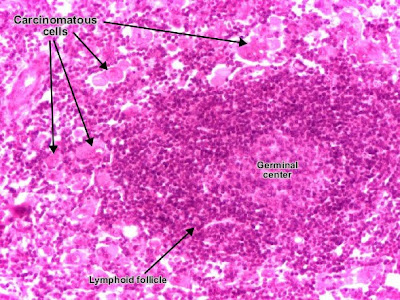
Histology and explanation of Carcinoma metastasis (lymph node)

Lymph node with carcinoma metastasis : clusters of tumor cells, atypical, with carcinomatous character.

Benefits of attending Top Medical Institution in the country


Becoming a medical professional is a challenging, daunting, and expensive path to follow. There are so many different paths that a person could follow – speaking in terms of different medical schools a person can attend – but they all lead to the same place. Regardless of the medical school that you choose, a person who graduates from medical school (and completes residency) will become a medical professional. After all, a medical professional from one school is still a medical doctor in another school. So, what’s the big deal in attending a top medical school, anyway?

For starters, reputation has a lot to do with contemporary society. Most people , including patients, have no idea what the rankings are of the top medical schools in the nation. As a result, even if you graduated from one of the Top Ten medical schools, your patients (and the general public) might not regard that accomplishment as being anything close to a doctor that graduated from a more popular prestigious medical school. In other words, how impressed an outsider is about your credentials, unfortunately, to a certain extent, on the popularity of the top medical school that you attended. From this perspective, usually possessed by a person that desires and relies on having graduated from a highly reputable medical school, then attending a top medical school might very well be a great decision.

Building on the idea of reputation, attending a top medical school has its perks. From the simple fact that the general public’s lack of knowledge about what the top medical schools are, people prefer more popular-named schools that those that are a little less known, however highly ranked that medical school may be.

But, what about the extremely lesser-known medical schools? Why would anyone want to attend medical schools that are not nationally ranked? For one, different people have different goals. If an individual wants to start up his or her own practice, then graduating from a top prestigious medical school is not as important an factor as for an individual who wants to be a plastic surgeon for famous celebrities. In the latter example, the patients that this particular doctor has might rely on his nationally ranked medical school credential to drive more patients to his office.

So, the main point is this: a medical professional from any medical school is still a medical professional. These doctors will more or less, depending on the specialty, make about the same annual salary. However, attending a top nationally ranked medical school can help satisfy a person’s reputation-related goals. It’s all a matter of perspective; and more importantly, it’s all a matter of personal preference.

Friday, February 25, 2011

Laparoscopic Herniotomy

Laparoscopic Herniotomy

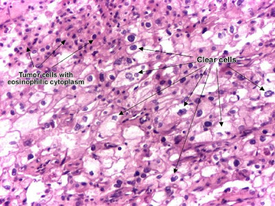
Histology and explanation of Renal cell carcinoma (Grawitz tumor)

Histology and explanation of Renal cell carcinoma (Grawitz tumor)
Renal clear cell carcinoma (Grawitz tumor) is a malignant epithelial tumor resulted from proliferation of tubule cells. Tumor cells form cords, papillae, tubules or nests, and are atypical, polygonal and large. Because these cells accumulate glycogen and lipids, their cytoplasm appears "clear", lipid-laden, the nuclei remain in the middle of the cells, and the cellular membrane is evident. Some cells may be smaller, with eosinophilic cytoplasm, resembling normal tubular cells. The stroma is reduced, but well vascularized. The tumor grows in large front, compressing the surrounding parenchyma, producing a pseudocapsule.

Histology Game: Histology Survey


Histology Survey Game

Instructions: In order to play the Histology Survey Game, you need a browser that supports flash. Make sure the sound to your computer is on.


Wednesday, February 23, 2011

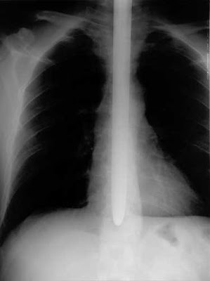
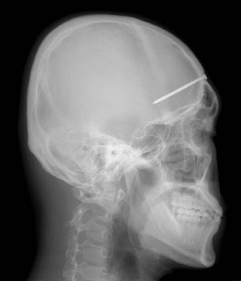
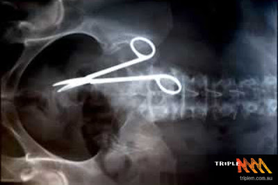
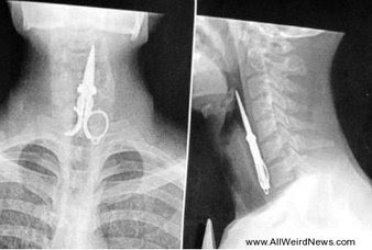
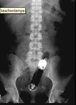
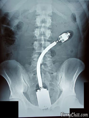
6 Most Funny and weird X-rays

6 Most Funny and weird X-rays


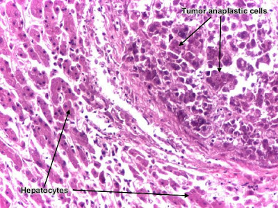
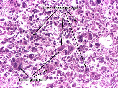
Histology and explanation of Poorly differentiated hepatocellular carcinoma

Histology and explanation of Poorly differentiated hepatocellular carcinoma
Hepatocellular carcinoma, poorly differentiated (photo - upper right), developed on liver cirrhosis. This malignant epithelial tumor consists in tumor cells, discohesive, pleomorphic, anaplastic, giant. The tumor has a scant stroma and central necrosis because of the poor vascularization. In well differentiated forms, tumor cells resemble hepatocytes, form cords and nests, and may contain bile pigment in cytoplasm.

Histology and explanation of Poorly differentiated hepatocellular carcinoma

Tuesday, February 22, 2011

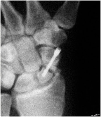
5 Most Funny and weird X-rays

5 Most Funny and weird X-rays


Most abnormal medical test results: A person having Blood alcohol of 0.55% but still alive


Victoria:
Na 174 (I think) in a very crispy elderly person. pCO2 of 20 in someone with muscular dystrophy and too much morphine on board.

Laura:
I've seen a Na of 107 before, highest K Ive seen is 8, highest Creatinine just under 3,000 and a Trop T of 17.

Cara:
wbc 55
neutro 52
a young women with atypical pneumonia.

Will:
I saw a guy in the royal with a trop T of 35, worse still it was about his fifth MI in six months, and the lowest Trop was 9, plus he didnt have renal failure contributing to the tropnin count.

I had a lady in AE who's sample was so lipaemic it was unmeasurable, we literally broke two of the labs very expensive UE analysers whcih caused chaos for the whole night... I remember taking the sample, there was about a half centimeter of fat visible floating on the top.

Tim:
Troponin of 960... 2 weeks post CABG... vein graft to LCx clotted off, clot propogated back up to the LM, then down the LAD... no renal failure... yes nine hundred and sixty.
Didn't survive...

Laura:
Neutrophil count of 0 in a patient with no known haematological problems, scared the bejeesus out of me as a a new house officer.

Tim:
Blood alcohol of 0.55%... chronic alcoholic...
The police didn't believe us that someone could be alive with a blood alcohol over 0.3, so we faxed the result to them... The patient was awake, alive and swearing at us.
The legal limit for driving in Australia is 0.05%.

Suzanne:
Our labs stop at the following -
K >10
Trop I >50
I've seen them both (different patients), and they survived

Other exciting ones...
Na 176 (dementia, NH resident)
Urea 72, Creatinine 860 (obstructed...)
Platelets 1 (ITP)
WCC 110 (CLL)
ABG pH 6.79 (DKA)


Please do share your experiences by writing a comment or sending an email at mdblogger20@gmail.com.

Histology Game: Histology Game Show (a histology quiz game)


Histology Game Show
(a histology quiz game)


Instructions: In order to play Histology Game Show (a histology quiz game), you need a browser that supports flash. If you have sound, turn it on.

The object of this histology quiz game is to get as close to $2400 as possible by correctly answering histology questions.

Questions are taken from the Histology Test Bank. Detailed explanations can be found in the Histology Test Bank.


Monday, February 21, 2011

Histology and explanation of Moderately differentiated adenocarcinoma (colon)

Histology and explanation of Moderately differentiated adenocarcinoma (colon)
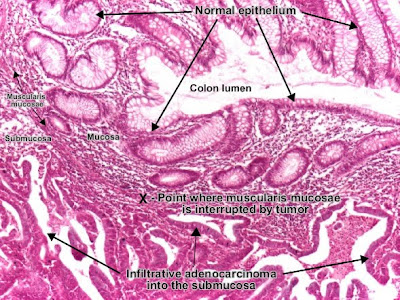
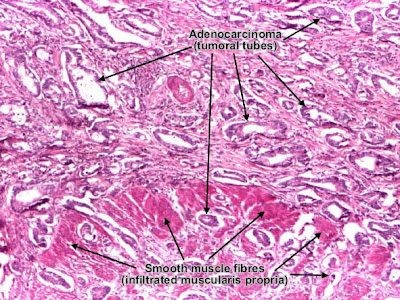
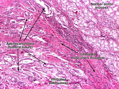
Adenocarcinoma is a malignant epithelial tumor, originating from glandular epithelium of the colorectal mucosa. It invades the wall, infiltrating the muscularis mucosae, the submucosa (photo) and thence the muscularis propria. (Notice the end-point of muscularis mucosae. At left from this point, muscularis mucosae is continuous. At right from this point, muscularis mucosae is destroyed by tumor cells invasion.) Tumor cells describe irregular tubular structures, harboring stratification, multiple lumens, reduced stroma ("back to back" aspect). Depending on glandular architecture, cellular pleomorphism and mucosecretion of the predominant pattern, adenocarcinoma may present 3 degrees of differentiation: well, moderate and poorly differentiate.

Sunday, February 20, 2011

Histology GameFling the Teacher (a histology quiz game)


Histology GameFling the Teacher (a histology quiz game)
Instructions: In order to play Fling the Teacher (a histology quiz game), you need a browser that supports flash. If you have sound, turn it on.

In order to win Fling the Teacher (a histology quiz game), you must correctly answer 15 histology questions in a row. If you miss a histology question, this histology quiz game ends. You have three options for help. You can ask a histology expert, you can see what answer other histology students voted for, or you can eliminate two incorrect answers. Each help option may be used only once.

Questions are taken from the Histology Test Bank. Detailed explanations can be found in the Histology Test Bank.


Histology and Explanation of Gastric carcinoma, intestinal type

Histology and Explanation of Gastric carcinoma, intestinal type

Gastric adenocarcinoma is a malignant epithelial tumor, originating from glandular epithelium of the gastric mucosa. According to Lauren classification, gastric adenocarcinoma may be: intestinal type, diffuse type and mixed type. Gastric adenocarcinoma, intestinal type. Tumor cells describe irregular tubular structures, with stratification, multiple lumens surrounded by a reduced stroma ("back to back" aspect). The tumor invades the gastric wall, infiltrating the muscularis mucosae, the submucosa and thence the muscularis propria. Often it associates intestinal metaplasia in adjacent mucosa. Depending on glandular architecture, cellular pleomorphism and mucosecretion, adenocarcinoma may present 3 degrees of differentiation : well (photo), moderate and poorly differentiate.

Histology and Explanation of Gastric carcinoma, intestinal type

A drug Alternative to alcohol - BBC video

Part-1:
Psychologist and addiction expert Dr John Marsden takes part in a radical experiment as he trials an non-addictive drug alternative to alcohol in this fascinating clip from Horizon.


Part-2:
A single injection reverses the 'drug' alternative to alcohol and Dr John Marsden is left to reflect on whether this is indeed be the way forward in reducing alcohol dependency in future generations.


Saturday, February 19, 2011

Histology and Explanation of Basal cell carcinoma

Histology and Explanation of Basal cell carcinoma

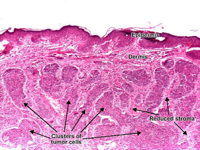
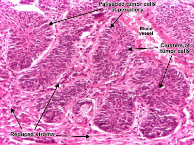
Basal cell carcinoma is a malignant epithelial tumor arising only in skin, from the basal layer of the epidermis or of the pilosebaceous adnexa. Tumor is represented by compact areas, well delineated and invading the dermis, apparent with no connection with the epidermis. Tumor cells resemble normal basal cells (small, monomorphous) are disposed in palisade at the periphery of the tumor nests, but are spindle-shaped and irregular in the middle. Tumor clusters are separated by a reduced stroma with inflammatory infiltrate.

Histology and Explanation of Basal cell carcinoma

Friday, February 18, 2011

Appendectomy: Surgical Removal of Appendix

Appendectomy: Surgical Removal of Appendix

Thursday, February 17, 2011

Histology game: Histology Tic-Tac-Toe: Bone

Histology game: Histology Tic-Tac-Toe: Bone

Instructions: In order to play histology tic-tac-toe, you need a browser that supports flash. Make sure the sound to your computer is on.


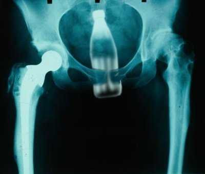
5 most funny and weird X-rays

5 most funny and weird X-rays


Pepsi-cola and Coca-Cola are found to contain carcinogens 2-methylimidazole and 4-methylimidazole


An ingredient used in Coca-Cola and Pepsi is a cancer risk and should be banned, an influential lobby group has claimed. The concerns relate to an artificial brown colouring agent that the researchers say could be causing thousands of cancers.

‘The caramel colouring used in Coca-Cola, Pepsi, and other foods is contaminated with two cancer-causing chemicals and should be banned,’ said the Center for Science in the Public Interest (CSPI), a health lobby group based in Washington, DC.

‘In contrast to the caramel one might make at home by melting sugar in a saucepan, the artificial brown colouring in colas and some other products is made by reacting sugars with ammonia and sulphites under high pressure and temperatures.

‘Chemical reactions result in the formation of two substances known as 2-MI and 4-MI which in government-conducted studies caused lung, liver, or thyroid cancer or leukaemia in laboratory mice or rats.’ America’s National Toxicology Program says that there is ‘clear evidence’ that both 2-MI and 4-MI are animal carcinogens, and therefore likely to pose a risk to humans.

The executive director of the CSPI, Michael F Jacobson, has petitioned America’s food regulator, the Food & Drug Administration, to take action.He said: ‘Carcinogenic colourings have no place in the food supply, especially considering that their only function is a cosmetic one.’

Mr Jacobson said the name ‘caramel colouring’ does not accurately describe the additives, explaining: ‘It’s a concentrated dark brown mixture of chemicals that simply does not occur in nature.

He added that while regular caramel could not be described as healthy, ‘at least it is not tainted with carcinogens’.

U.S. regulations distinguish between four types of caramel colouring, two of which are produced with ammonia and two without it. The CSPI wants the two made with ammonia to be banned and has received backing from five prominent cancer experts, including several who have worked at the National Toxicology Program.

The type used in colas and other dark soft drinks is known as Caramel IV, or ammonia sulphite process caramel. Caramel III, which is produced with ammonia but not sulphites, is sometimes used in beer, soy sauce, and other foods.

The CSPI admitted that any risk associated with consumption of the chemicals would be extremely small. It said the ten teaspoons of sugar found in a can of regular cola would be more of a health problem.

However, it argued the levels of 4-MI in the tested colas still may be causing thousands of cancers in the U.S. population alone.
Histology Game: Histology Anagrams


Instructions: In order to play Histology Anagrams, you need a browser that supports flash. If you have sound, turn it on. To play Histology Anagrams, unscramble the histology words. You have half a minute to unscramble each histology word.


Diabetic Retinopathy Animation


Diabetic Retinopathy Animation

Wednesday, February 16, 2011

Each day the average heart beats 100,000 times and pumps about 2000 gallons of blood


The normal heart is a strong, muscular pump a little larger than a fist. It pumps blood continuously through the circulatory system. Each day the average heart "beats" (expands and contracts) 100,000 times and pumps about 2,000 gallons of blood. In a 70-year lifetime, an average human heart beats more than 2.5 billion times.

The circulatory system is the network of elastic tubes that carries blood throughout the body. It includes the heart, lungs, arteries, arterioles (ar-TE're-olz) (small arteries), and capillaries (KAP'ih-lair"eez) (very tiny blood vessels). These blood vessels carry oxygen- and nutrient-rich blood to all parts of the body. The circulatory system also includes venules (VEN' yoolz) (small veins) and veins. These are the blood vessels that carry oxygen- and nutrient-depleted blood back to the heart and lungs. If all these vessels were laid end-to-end, they'd extend about 60,000 miles. That's enough to encircle the earth more than twice.

The circulating blood brings oxygen and nutrients to all the body's organs and tissues, including the heart itself. It also picks up waste products from the body's cells. These waste products are removed as they're filtered through the kidneys, liver and lungs.

What is the heart's structure?
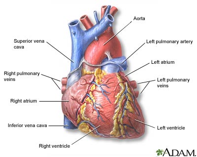
The heart has four chambers through which blood is pumped. The upper two are the right and left atria. The lower two are the right and left ventricles. Four valves open and close to let blood flow in only one direction when the heart beats:

  • The tricuspid valve is between the right atrium and right ventricle.
  • The pulmonary or pulmonic valve is between the right ventricle and the pulmonary artery.
  • The mitral valve is between the left atrium and left ventricle.
  • The aortic valve is between the left ventricle and the aorta.

Each valve has a set of flaps (also called leaflets or cusps). The mitral valve has two flaps. The others have three. Under normal conditions, the valves let blood flow in just one direction. Blood flow occurs only when there's a difference in pressure across the valves that causes them to open.

How does the heart pump blood?
A heart's four chambers must beat in an organized manner. This is governed by an electrical impulse. A chamber of the heart contracts when an electrical impulse moves across it. Such a signal starts in a small bundle of highly specialized cells in the right atrium — the sinoatrial (SI'no-A'tre-al) node (SA node), also called the sinus node. A discharge from this natural "pacemaker" causes the heart to beat. This pacemaker generates electrical impulses at a given rate, but emotional reactions and hormonal factors can affect its rate of discharge. This lets the heart rate respond to varying demands.

Wireless phones as a cause of brain cancers


Appearing in the Journal of Computer Assisted Tomography (Nov/Dec 2010), this study attempts to filter data from old studies by limiting analysis to long-term use, re-defining what can be called an "exposure," and modifying which tumors can be counted as occuring in the presence of a wireless phone exposure. See Science Daily, Jan 19, for a journalistic view of the original publication.

This is a great example of fiddling with the data to support your conclusion. Not that they don't have a rationale for modifying others research data, but there's an inherent danger to modifying end points of an experiment after the fact. If you design an experiment to look at the occurrence of result A, then after the experiment is over you decide to look back at the data for a different result B (for which the experiment was not designed) bias and error can intrude. Not always, though.

The whole question of whether wireless phones (expanding the definition to include both cellular and home wireless phones) are a risk for cancer is undecided because the dozens of studies can all be criticized. If you go by raw numbers, there are just as many studies that conclude "yes there is a risk" as there are "no there's no risk."

The authors admit that their study is not definitive, but argue that the possibility of harm is sufficient to warrant economically feasible methods to reduce exposure while further studies are done. I don't have a problem with the "precautionary principle" where concern is established, but I doubt that the recommendations the authors make will be implemented: for instance, using "tube" type earpieces instead of wired ear plugs (the wired version radiates energy from the phone and can act as an antenna for other exposure. I'm not sure that's been demonstrated to occur).
This article does a couple of useful things. It focuses more on long-term use, assuming a cumulative effect of exposure with phone use. And, they point out that industry-funded studies have been more likely to show no hazard than independently funded studies.

But, if you're the kind of person who spends several hours on the phone every day, it's possible that you have other....uh...issues. (Does phone addiction exist?)

Histology and explanation of Squamous cell carcinoma (skin)

Histology and explanation of Squamous cell carcinoma (skin)

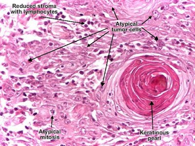
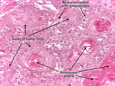
Squamous cell carcinoma is a malignant epithelial tumor which originates in epidermis, squamous mucosa or areas of squamous metaplasia. In skin, tumor cells destroy the basement membrane and form sheets or compact masses which invade the subjacent connective tissue (dermis). In well differentiated carcinomas, tumor cells are pleomorphic/atypical, but resembling normal keratinocytes from prickle layer (large, polygonal, with abundant eosinophilic (pink) cytoplasm and central nucleus). Their disposal tends to be similar to that of normal epidermis: immature/basal cells at the periphery, becoming more mature to the centre of the tumor masses. Tumor cells transform into keratinized squames and form round nodules with concentric, laminated layers, called "cell nests" or "epithelial/keratinous pearls". The surrounding stroma is reduced and contains inflammatory infiltrate (lymphocytes). Poorly differentiated squamous carcinomas contain more pleomorphic cells and no keratinization. (H&E, ob. x10)Squamous cell carcinoma (skin). Tumor cells transformed into keratinized squames form round nodules with concentric, laminated layers, called "cell nests" or "epithelial/keratinous pearls". (H&E, ob. X40)Histology and explanation of Squamous cell carcinoma (skin)

Top three reasons to drop out of Medical colleges


Medical life is a daunting experience. There are ups and downs, and everyone entering Medical school knows that; expects that. Every new medical school student has that dream of being called Doctor Somebody. However, how does one really know when enough is enough? How does one really know if the medical path is not the right path to follow… for them? Yes, it may be the right path for that guy, or that girl; but, is it really for you?

This article attempts to investigate three reasons that might be evident signs prompting you to drop out of medical school… now. It is in no way meant to be derogatory toward the medical profession, as it has its unparalleled benefits, both in salary and human reward. Instead, this article was written to provide some insight into three reasons, among many other reasons, why medical school might not be for certain people.

You should probably drop out of medical school if…

1) You plan on having a family during your “earlier” years. If you have always dreamed of raising your own family at a relatively young (in your twenties) age, then you might want to consider the “drop out” option of attending medical school. It definitely is possible to have your own family throughout your medical school years, but!… the more important question you should be asking yourself is how much time am I willing to sacrifice away from my family? Precious time spent away from one’s family, especially in the beginning years, is precious time you may never get back. Do you really want to be a “stranger” to your kids at this extremely important time in their lives?

2) You’re still in it just for the money. Yes, you may be able to keep up with the work that is required of medical school students. However, is being a doctor really what you have a burning desire to do for the rest of your life? Sure, the money is great – few other occupations are on par with the yearly salaries of medical professionals, but consider what the trade-off is. Are you willing to work hard during a great chunk of your life just for a little more green paper flowing in? Think about it for a little bit.

3) Your cherished relationships are suffering because of it. As a general rule in life, you should drop out of anything that does not bring you the happiness you deserve; the happiness you are meant to experience. Some medical students undergo much stress, but their perspective is clear: “I want to be a doctor, and if this is what it takes, nothing’s going to stop me.” However, if you fall into the category of those that wake up dreading the day, day after day; then, you probably should drop out of medical school.

It seems as though all the arrows to drop out of medical school point toward what makes a person happy.

As a matter of fact, it is not difficult to distinguish whether or not medical school is for you. All you have to do is be honest with yourself.

Do you really want to be a doctor?

Whether your answer is yes or no, you can find out right now if you just allow yourself to be completely honest.

If Yes, you can read few helpful tips that will help you succeed in medical life.

Histology game: Spin the Wheel

Instructions: In order to play Spin the Wheel (a histology quiz game), you need a browser that supports flash. Make sure the sound to your computer is on. Hit button in the center of the wheel to spin the needle. Answer the histology question that follows. Click Start to begin.


How to Take a Good Obstetrical History & Performing a Thorough Clinical Examination

HISTORY:

Demographical Details/Personal Profile:

1. Name

2. Age

3. Education

4. Occupation

5. Marital Status (if married, for how long? )

6. Residence

7. Date, time & mode of Admission

8. Which pregnancy?

Presenting Complaints:

1. Gestational amenorrhoea and its duration (in weeks)

2. Any other complaint

a. Labor pains

b. PROM

i. PROM at term

ii. PPROM

c. Vaginal bleeding (APH, PPH etc.)

d. Abdominal pain (other than labor pains)

e. Decreased/absent fetal movements

f. Seizures

g. HTN or Pre-eclampsia or Eclampsia (Symptoms)

h. DM or GDM (Symptoms)

i. Anemia (Symptoms)

j. Hyper/hypothyroidism (Symptoms)

k. Ischemic Heart Disease (Symptoms)

l. Valvular Heart Disease (Symptoms)

History of Present Pregnancy:

1. Planned/ Unplanned

2. Wanted/ Unwanted

3. Reaction to pregnancy

4. LMP (first day of last menstrual period)

5. Use of Contraception

6. Any spotting or bleeding

7. Specific things to ask in First Trimester

a. Confirmation of pregnancy, how and when?

b. Any symptoms of pregnancy (Nausea, vomiting etc.)

c. Urine/Serum Pregnancy test

d. Ultrasonography

e. Any antenal visit?

8. Specific things to ask in Second Trimester

a. When did patient start to feel fetal movements?

b. Any anomaly scan?

c. Any prenatal diagnosis? If yes, then what?

9. Specific things to ask in Third Trimester

a. Any CTG, USG or BPP done

b. Results of the above

c. Any findings on antenatal visits

d. Any hospital admission? If yes, then for what problems?

e. Fetal movements

f. Vaginal discharge (PROM)

g. Vaginal bleeding

Depending upon the other presenting complaint, following questions may be relevant to ask

1. Onset

2. Duration

3. Intensity/Severity

4. Aggravation and relieving factors 5. Associated features

Past Obstetrical History:

1. Number of children

2. Ages of all children

3. Mode of deliveries of all children

4. Gestational age of all children at the time of delivery (Term/Preterm etc.), present state of health (Alive/Died Later/Handicapped/Retarded)

5. Number of miscarriages and duration of gestation at those

6. Termination of pregnancy/pregnancies, At what gestation? For what reason?

Menstrual History:

1. Age of menarche

2. Cycle Regular or Irregular

3. Duration of Cycle

4. Quantity of Blood loss

5. Dysmenorrhoea

6. Intermenstrual Bleeding

7. Post coital Bleeding

8. Dyspareunia

9. LMP

10. Cervical Smear

Past Gynecological History:

1. Any previous gynecological problems

2. Any treatment for the same

3. Cervical Swab

Sexual History:

1. Age at first intercouse

2. Coital freuency

3. Any coital difficulty

4. Number of sexual partners

5. Number of sexual partners of the patient’s partner/husband

Past Medical History:

(Please refer to clinical methods of Internal Medicine for details.)

Past Surgical History:

(Please refer to clinical methods of Internal Medicine for details.)

Systemic Inquiry:

From CVS, CNS, RS, GIT, GUT & MSK

(Please refer to clinical methods of Internal Medicine for details.)

Family History:

About Infectious diseases, IHD, HTN, DM, Multiple Pregnancy, Gynecological and other malignancies

(Please refer to clinical methods of Internal Medicine for details.)

Social/ Socioeconomic/ Biosocial History:

1. Place of living

2. Type of family

3. Monthly Income

Personal History:

(Please refer to clinical methods of Internal Medicine for details.)

Drug History:

(Please refer to clinical methods of Internal Medicine for details.)

Treatment History:

(Please refer to clinical methods of Internal Medicine for details.)

Examination:

General Physical Examination:

(Please refer to clinical methods of Internal Medicine for details.)

Systemic Examination:

CNS (Please refer to clinical methods of Internal Medicine for details.)

CVS (Please refer to clinical methods of Internal Medicine for details.)

RS (Please refer to clinical methods of Internal Medicine for details.)

Abdominal Examination:

1. Inspection

2. Palpation

a. Assessment of fundal height

i. Palpatory method

ii. Measurement method

b. Assessment of fetus

i. First manoeeuvre (Fundal palpation)

ii. Second manoeuvre (Lateral palpation)

iii. Third manoeuvre (Pawlik’s grip; palpation of presenting part)

iv. Fourth manoeuvre (For attitude of fetal head & engagement of head in fifths; 5/5, 4/5, 3/5, 2/5, 1/5)

3. Ausculation (with pinard stethoscope for fetal heart sounds)

Pelvic Examination: (Generally not required, specially contraindicated in vaginal bleeding (APH) or vaginal discharge)

(Ensure Adequate Privacy)

1. Inspection

a. Hair Distribution

b. Labial Appearance

c. Episiotomy Scar etc.

d. Stress incontinence

e. Vaginal Discharge

2. Palpation

3. Speculum Examination (for vaginal discharge, to confirm/exclude PROM)

4. Vaginal Examination (Not indicated if woman not in labor)

Only done in two conditions;

i. Before labor (Bishop scoring)

ii. During labor (to assess progress of labor; Dilatation of cervix & length of cervix)

Provisional/Working Diagnosis + Risk Scoring (High/Low Risk):

Differential Diagnosis (es):

Investigations:

a. Routine

b. Specific

Definite Diagnosis:

Plan of Management:

SUMMARY:

Most abnormal medical test results: A person having Blood Sugar of >1200 & pH of 6.8 and he did not know he was diabetic


Will:
1) Alcoholic with refeeding syndrome - magnesium undetectable
2) COPD and CCF - ABG PaO2 3.2
3) TB peritonitis - White cells 86, CRP 1332
4) Octogenerian living on tinned soup - MCV 149.6, Hb 4.2, Folate 0.1
5) 3 days unrecognised DKA - Base XS -35, pH 6.69, pCO2 0.1 Lactate 21

Michael:
Hb 3.2 - last week, bleeding DU. Still conscious!

Mark:
Creatine Kinase of 326,000 in a 22 year old student who'd suffered prolonged seizures following overdose of a TCA - did very well on intensive IV fluid therapy on HDU and went home reasonably quickly thereafter.

Adegbenga:
1. H/H 1.2/3.5 walked into ED with swollen feet. Died following 2 units of blood over rapid transfuser, Clever ED!!

2. Blood Sugar of >1200 and pH of 6.8, Na of 177 in the same Px with DKA and he did not know he was diabetic

Tom:
Sickler mid-crisis - hB undetectable by haemocue or ABG. Lab result confirmed haemoglobin of 0.5. He was still awake, alert, and refusing treatment (violently).

Clare:
Urea 70 Creat 1000 K 7.something, after 3/52 urinary retention.

Patricia:
Ur 68 Cr 2100 K7.5 generally not feeling well but only came to hospital because wife wouldn't go by herself in the ambulance and thought he'd get checked out in A&E too. He had bilateral hydronephrosis due to renal stones. There was another lady the same week with a K of 10 who survived.


Please do share your experiences by writing a comment or sending and email at mdblogger20@gmail.com.

Tuesday, February 15, 2011

Top 12 rules to live a Happy and Successfull life


  • Know what you can and want to do in life.
  • Set goals for yourself and work hard to achieve them.
  • Strive to have fun every day.
  • Use your creativity as a means of expressing your feelings.
  • Be sensitive in viewing the world.
  • Develop a sense of confidence.
  • Be honest with yourself and with others.
  • Know that the more you give the more you will receive.
  • Believe in yourself and your dreams will come true.
  • One day you will see that it all has finally come together.
  • What you have always wished for has finally come to be.

Robotic Surgery Of Infertility video and description

Robotic Surgery Of Infertility

How to Take a Good Gynecological History & Performing a Thorough Clinical Examination

HISTORY:

Demographical Details/Personal Profile:

1. Name

2. Age

3. Education

4. Occupation

5. Marital Status (if married, for how long? )

6. Residence

7. Date (and time) of Admission

8. Mode of Admission

Presenting Complaints:

Common complaints encountered in a gynecological patient are

1. Abnormal Menstruation

a. Pattern (Regular/Irregular)

b. Amount of blood loss

c. Passage of blood clots

d. Duration of menstruation

e. Intermenstrual Bleeding

f. Post coital Bleeding

2. Bleeding in early pregnancy i.e. before 24 weeks of gestation (Abortion/Miscarriage)

3. Something coming out of vagina

4. Pelvic Mass

5. Vaginal Discharge – amount, colour, odour, presence of blood, relation to periods

6. Pelvic pain – site, nature and relation to periods. Anything that aggravates or relieves the pain

History of Present Illness:

Depending upon the presenting complaint, following questions may be relevant to ask

1. Onset

2. Duration

3. Intensity/Severity

4. Aggravation and relieving factors

5. 5. Associated features

Menstrual History:

1. Age of menarche

2. Cycle Regular or Irregular

3. Duration of Cycle

4. Quantity of Blood loss

5. Dysmenorrhoea

6. Intermenstrual Bleeding

7. Post coital Bleeding

8. LMP

Past Gynecological History:

1. Any previous gynecological problems

2. Any treatment for the same

3. Cervical Swab/ Cervical Smear

Past Obstetrical History:

1. Number of children

2. Ages of all children

3. Mode of deliveries of all children

4. Gestational age of all children at the time of delivery (Term/Preterm etc.) + Presence state of health

5. Number of miscarriages and duration of gestation at those

6. Termination of pregnancy/pregnancies, At what gestation? For what reason?

Sexual and Contraceptive History:

1. Age at first intercouse

2. Coital freuency

3. Dyspareunia

4. Any coital difficulty

5. Number of sexual partners

6. Number of sexual partners of the patient’s partner/husband

7. Use of contraception? Type of contraception used?

Past Medical History:

(Please refer to clinical methods of Internal Medicine for details.)

Past Surgical History:

(Please refer to clinical methods of Internal Medicine for details.)

Systemic Inquiry:

From CVS, CNS, RS, GIT, GUT & MSK

(Please refer to clinical methods of Internal Medicine for details.)

Family History:

About Infectious diseases, IHD, HTN, DM, Multiple Pregnancy, Gynecological and other malignancies

(Please refer to clinical methods of Internal Medicine for details.)

Social/ Socioeconomic/ Biosocial History:

1. Place of living

2. Type of family

3. Monthly Income

Personal History:

(Please refer to clinical methods of Internal Medicine for details.)

Drug History:

(Please refer to clinical methods of Internal Medicine for details.)

Treatment History:

(Please refer to clinical methods of Internal Medicine for details.)

Examination:

General Physical Examination:

(Please refer to clinical methods of Internal Medicine for details.)

Systemic Examination:

Abdominal (Please refer to clinical methods of Internal Medicine for details.)

CNS (Please refer to clinical methods of Internal Medicine for details.)

CVS (Please refer to clinical methods of Internal Medicine for details.)

RS (Please refer to clinical methods of Internal Medicine for details.)

Pelvic Examination: (Not expected from Final Year students.)

(Ensure Adequate Privacy)

1. Inspection

a. Hair Distribution

b. Labial Appearance

c. Episiotomy Scar etc.

d. Stress incontinence

e. Vaginal Discharge

2. Speculum Examination

a. Inspection of vaginal walls (Atrophic/Healthy/Hyperemic/Growth/Ulcers/Polypi etc.)

b. Appearance of cervix (Ectopy/Suspicious Polypi)

c. Vaginal discharge

3. Bimanual Examination/Palpation

a. Consistency of cervix

b. Cervical os

c. Uterine consistency, size, shape and mobility

d. Uterus retroverted or anteverted

e. Adnexal Mass

f. Adnexal Tenderness

g. Pouch of Douglas

Provisional/Working Diagnosis:

Differential Diagnosis (es):

Investigations:

a. Routine

b. Specific

Definite Diagnosis:

Plan of Management:

SUMMARY:

Diabetic Nephropathy Animation


Diabetic Nephropathy Animation

Monday, February 14, 2011

Schwartz's Principles of Surgery Available for iPhone


Schwartz's Manual of Surgery has become the resource of choice for both practicing physicians, residents, and even surgical clerkship students who are interested in a comprehensive, yet concise on-the-job manual detailing the most current practices and trends.

This edition reflects the most extensive revisions ever with a greater focus on surgical techniques and a superb array of new visuals including more tables, charts, highlighted topics throughout. New to this edition: staging tables for cancer, anesthesia guidance, pre-and-postoperative management of the surgical patient, and much more.

View On itunes