This is default featured slide 1 title
Go to Blogger edit html and find these sentences.Now replace these sentences with your own descriptions.
This is default featured slide 2 title
Go to Blogger edit html and find these sentences.Now replace these sentences with your own descriptions.
This is default featured slide 3 title
Go to Blogger edit html and find these sentences.Now replace these sentences with your own descriptions.
This is default featured slide 4 title
Go to Blogger edit html and find these sentences.Now replace these sentences with your own descriptions.
This is default featured slide 5 title
Go to Blogger edit html and find these sentences.Now replace these sentences with your own descriptions.
Thursday, March 31, 2011
Histology and Explanation of Coronary atherosclerosis - fibro fatty plaque

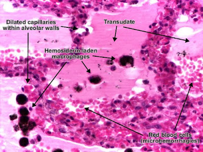
Histology and explanation of Passive congestion (Passive hyperemia) (lung)
Active hyperemia (congestion) is a result of arteriolar distension (e.g., skeletal muscle activity, inflammation, local neuro-vegetative reaction).

Passive hyperemia (congestion), also termed stasis, is a consequence of an impaired venous drainage (heart failure, compression or obstruction of veins), followed by dilatation of venules and capillaries.

Etiology of passive congestion of the lung : chronic left heart (ventricular) failure.
Alveolar walls are thickened due to dilated capillaries. Alveolar lumens are filled with transudate (amorphous, eosinophilic and homogenous) which replaced the air, red blood cells (microhemorrhages) and hemosiderin-laden macrophages (also called "heart failure cells").
With progression, interstitial fibrosis may appear and, together with hemosiderin pigmentation, generates the aspect of "brown induration". Extensive fibrosis leads to intrapulmonary hypertension.
Passive congestion of the lung. Hemosiderin-laden macrophages contain in cytoplasm hemosiderin pigment (brown, granular), resulted from destruction of red blood cells in alveolar lumen.
Perls reaction is useful in distinguishing hemosiderin pigment from anthracotic (carbon) pigment : trivalent iron from hemosiderin stains in green-blue, while anthracotic pigment remains dark-brown and is mainly located perivascular.
Friday, March 18, 2011
Histology and Explanation of Pulmonary Edema

Edema represents the accumulation of excess liquid in the interstitial (extracellular) spaces of a tissue or in pre-existing cavities. It may affect any organ, but most often it appears in : subcutaneous tissues, lung and brain.
According to the etiology, edema may be localized (in inflammation or in impaired venous drainage) or systemic (in right heart failure or in nephrotic syndrome). A generalized and severe edema is called anasarca.
Accumulation of transudate or non-inflammatory fluid (effusions) in body cavities :
* Peritoneal cavity - ascites
* Pleural cavity - hydrothorax
* Pericardial cavity - hydropericardium
Pulmonary edema
Etiology of pulmonary edema : acute left heart (ventricular) failure, pulmonary failure in syndrome of adult respiratory distress, pulmonary infections and hypersensitivity reactions.

Pulmonary edema. Alveolar walls are thickened due to acute distention of capillaries and interstitial edema. Alveolar lumen is filled with transudate (pale-eosinophilic, finely granular), a liquid which replaces the air.
Thursday, March 17, 2011
Histology and Explanation of Benign microcystic teratoma of the ovary
Wednesday, March 16, 2011
Download Clinical Nutrition: Enteral and Tube Feeding, 4th ed. 2005 Free
Rolando Rolandelli, Robin Bankhead, Joseph Boullata, Charlene Compher
ISBN-10: 0721603793
ISBN-13: 978-0721603797
This definitive reference presents the most comprehensive, clinically relevant coverage of nutrition in enteral and tube feeding. The New Edition has been completely revamped by a multidisciplinary editorial team to reflect all of the latest technology and nutritional knowledge, as well as the new, collaborative nature of contemporary clinical practice. Plus, a new bonus CD-ROM delivers self-assessment questions and answers, and a downloadable image collection of illustrations from the book.
* Delivers 21 brand-new chapters that address recent ASPEN clinical guidelines regarding pharmacotherapeutic issues and enteral formulations, including fluids and electrolytes · genetics · pre-, pro-, and synbiotics · food safety · regulatory issues · and more.
* Explores the unique enteral nutrition needs of specific patient populations such as pregnant and lactating women · children · adolescents · transplant candidates · cancer patients · obese patients · patients with kidney disorders · and diabetic patients.
* Features new editorial viewpoints representing surgery, nursing, pharmacy, and nutrition/dietetics, reflecting the new, multidisciplinary nature of the field.
* Offers a new bonus CD-ROM containing review questions and answers and more.
* Presents a new, more user-friendly internal
* An excellent study preparation tool for Nutrition Support Certification.
How to Treat Diabetes Mellitus

Type one diabetes is when cells in the pancreas have been destroyed which causes a lack of insulin being produced. There may be many reasons for this to happen. An infection caused by a particular virus or bacteria, exposure to chemical toxins in food, exposure to cow milk as a really young infant are three ideas of causes of why diabetes happens in a person.
Type 2 diabetes develops when the receptors on cells which respond to insulin stop being stimulated by it, commonly referred to as insulin resistance. Type 2 diabetes would even develop when there's just not enough insulin available or the insulin available is abnormal in some way.
Diabetes Help:
You may find many different medications that are available for diabetes treatment. It is important that if you have symptoms of diabetes, you need to get some help from your physician. This is not a disease which can be taken care of on your own. Once you are diagnosed with diabetes your physician will prescribe diabetes treatments that would be specifically designed to meet your requirements.
Individualized diabetes treatments are important because each person is different. When looking to control blood sugar levels it will take special diabetes treatments designed with your particular requirements in mind. Few people will need to take insulin in order to control their diabetes, whilst others could make use of a simple diet and exercise program to control their diabetes.
Diet and exercise are the number one diabetes treatments which would be prescribed. It would become very vital to watch what you eat when you are diagnosed with the disease. If you are struggling with your diet or exercise program there are diabetes help programs available for you to call upon.
There are increasingly more individuals being diagnosed with diabetes in the United States. Diabetes help programs are developed to give people the support they need to completely understand their disease and the way to deal with it. Learning to live with diabetes 1 or 2 could be difficult, as it would need a major life-style change for several people. Reaching out for aid is vital as coping with diabetes can become rather hard at times.
The primary thing to remember is to follow your doctor's orders. The medication that's prescribed to you would only assist if it is taken correctly. If you feel that the treatment plan you're presently on is not helping its important to discuss it with your doctor and get the regimen changed until it fully meets your needs.
Helpfull Tips for you to survive in a Medical school
The volume of material to be learned in medical school is greater than you have experienced before. Feeling helpless and pressured is normal. Exhaustion, can at times be overwhelming. But you are not alone.
1-Take care of yourself first:
Taking care of yourself is not selfish act bu...t a fact that can help your personal and professional growth. Many of us do not come close to practicing what we teach patients, we need to follow our own advice regarding health.
2-Stay Connected:
An important task in medical school is to build and maintain social connections. Life should not end just because you are in medical school. Don’t abandon your spouse, partner, lover, friends, or family. Their love and support for you is potentially the most important factor for success.
3- Find Friends in Class:
This is an important thing you can do to ensure your survival and enjoyment of medical school.
4-Take Care of Your Body:
Fast food, vending machines, too little sleep, and too much sitting may all wreak havoc on your health. Too much caffeine can cause anxiety, interfere with sleep and cause fatigue. Cooking takes time, but shared meal preparation and finding more healthy snacks are important.
5-Exercise your body:
Physical activity improves bodily function and helps clear the mind. Exercise can be both social and fun. Find enjoyable activities, particularly those that include companions.
6-Plan Your Career:
Look for what you like and do best. The life of a physician can be lived in many ways. Give yourself time during medical school to explore who you are and what you want. The choice must be yours.
7-Plan yourself:
There are two types of med students: those who study for several hours every day, and those who cram like crazy in the days before a test. We recommend the former.
8-Take a break:
...Like everyone else, med students need time to veg out, reconnect with friends, or catch up on sleep. Set aside a few hours each week to relax and enjoy yourself, whatever that means to you.
9-Get help when you need it:
If you are are feeling overwhelmed by stress, discuss the situation with your dean. He or she will be able to advise you on the best course of action
10-Don’t sweat the small stuff:
You would not have been accepted if you didn’t have what it takes to succeed. Learning how to perform under difficult and demanding circumstances is an important part of becoming a doctor.
Tuesday, March 15, 2011
Download Publication Manual of the American Psychological Association, 6th ed. 2010 free
American Psychological Association
ISBN-10: 1433805618
ISBN-13: 978-1433805615
The Publication Manual of the American Psychological Association" is the style manual of choice for writers, editors, students, and educators in the social and behavioral sciences. It provides invaluable guidance on all aspects of the writing process, from the ethics of authorship to the word choice that best reduces bias in language. Well-known for its authoritative and easy-to-use reference and citation system, the Publication Manual also offers guidance on choosing the headings, tables, figures, and tone that will result in strong, simple, and elegant scientific communication. The sixth edition offers new and expanded instruction on publication ethics, statistics, journal article reporting standards, electronic reference formats, and the construction of tables and figures. The sixth edition has been revised and updated to include: new ethics guidance on such topics as determining authorship and terms of collaboration, duplicate publication, plagiarism and self-plagiarism, disguising of participants, validity of instrumentation, and making data available to others for verification; new journal article reporting standards to help readers report empirical research with clarity and precision; simplified APA heading style to make it more conducive to electronic publication; updated guidelines for reducing bias in language to reflect current practices and preferences, including a new section on presenting historical language that is inappropriate by present standards; new guidelines for reporting inferential statistics and a significantly revised table of statistical abbreviations; and, new instruction on using supplemental files containing lengthy data sets and other media.
Treatment choices for Breast Cancer
In the healthcare world today, the treatment for breast cancer involves many kinds of approaches. The initial analysis and clinical work up is the guide towards the type of remedy you will undergo.You can find some requirements which your remedy depends like the cellular composition of the cancer, the dimensions of the growth, and the phase of your hormonal status. Your general health status also plays a role in deciding the type of treatment. This information will take you through several from the latest treatments on breast cancer.
Generally there is really a good deal happening in your tissue. In the event that the cells that are developing out of control are normal cells, the growth is not cancerous. However, if the tissues which are developing out of control are abnormal and do not perform like the body's usual cells, the tumor is malignant.
Quite a few individuals choose surgical treatment. Surgical treatment and radiation therapy are most effective when the growth is localized towards the breast and can be easily removed. Benign tumors and small sized tumors are treated in this manner. The newest treatments on breast most cancers include improvements in chemotherapy, hormone treatment. These are utilized in advanced phases of cancer when the growth is no longer confined just to the breast.
The actual reproduction of cells becomes imbalanced. Breast cancer is if your cells begin to grow and reproduce out of control, which creates an assortment of cells called a tumor. However, just because you have a unknown growth within the breast doesn't mean it needs to be dangerous.
One option is to test is opting for different kinds of surgery. Breast cancer surgery is aimed at removing the bulk from the cancer tissue. Better surgical options have emerged which give patients the chance to undergo reconstruction surgery after a mastectomy. Surgery may also be within the breast or lymph node which may be found during a diagnosis. Newest remedies on this topic have medical choices which include a prophylactic mastectomy or perhaps a removal of the sex gland in a few instances.
The source from the names for each cancer is rather easy. They are named after the part from the body from which they develop. Breast cancer stems within the breast tissue. Like other cancerous tissues, it can invade and grow into the tissue surrounding the breast. Additionally , it may pass through to other parts of the human body and form brand new growths. This is called metastasis and can be very dangerous
There are many dangerous but sometimes effective methods to combat cancer. Chemotherapy is considered systemic therapy as it targets cancerous cells within the entire body. It can also be supplemented by a bone marrow or stem cell transplant that is beneficial in most cases where the immune system is severely compromised. Stem cell treatment offers incredible possibilities in cases like breast cancer which have the potential to turn into any of the body's cells and replicate at a faster pace to fight this lethal disease.
Monday, March 14, 2011
Baby Boy Cries With One Eye Open. WHAT'S YOUR DIAGNOSIS?
HISTORY
This 1-week-old baby boy was brought for his first newborn visit. The parents were concerned that when he cried, the left side of his face “does not move”.
He was born at full term via normal vaginal delivery to a primigravida without complications. Weight was 3.47 kg. Apgar scores, 9 and 9 at 1 and 5 minutes. Pregnancy and prenatal course were uneventful. Mother denied history of uterine tumors, multiple gestation, or polyhydramnios. No family history of neurological disorders or genetic syndromes.
PHYSICAL EXAMINATION
Vital signs normal. Weight, 3.51 kg; length, 49.5 cm; and head circumference, 34 cm. At rest, the infant's facial features appeared symmetric (A), with no bruising or obvious trauma.

When he cried, the lower lip on the left side did not depress, the left eyelids remained open, the forehead and eyebrow did not wrinkle, and the nasolabial fold on the left was flat (B).

He had normal suck and swallow reflexes and did not drool.
Mild positional plagiocephaly was noted in the right parietal area. Anterior fontanelle open, 2 × 2 cm, and pulsatile. Red reflexes intact bilaterally, with no obvious retinal hemorrhages. Two small subconjunctival hemorrhages noted in the right eye were coalescing around the iris and appeared to be of no clinical significance.
WHAT'S YOUR DIAGNOSIS?
What are the challenges of residency in medicine and how to cope with them

Your graduation from medical school does not mark the end of your education. After earning an MD, you will spend several years as a resident in a teaching hospital. Residency can be a grueling experience. When it comes to the hospital environment, you're at the bottom. On the flip side, you will work directly with patients and gain mastery of a specific specialty.
The residency can last anywhere from three to eight years, depending on your specialty. You’ll learn almost everything you need to know about your area of specialty during this time. Expect exhaustion to be a constant factor. Typical work weeks range from 50 to 80 hours, and residents spend several nights a week on-call. The first three to five months of residency will be the hardest, as your body adjusts to the lack of sleep, surplus of work, and constant reprimanding that comes with the territory.
As the residency progresses, you will be given more responsibility and make more decisions independently of your attending physician. Once your residency is over, you’ll finally have the knowledge and experience to practice medicine on your own. Some doctors pursue a fellowship, Others join a hospital, clinic start their own private practice
Saturday, March 12, 2011
Pregnancy Test Made easy by USB Digital Pregnancy Test

- Pee on the stick, specifically the absorbent test strip at one end.
- Remove the cap from the other stick end, to reveal the USB connector.
- Pop the device in your computer.
- When you’re done with the test result, pop it out to your computer and change the strip. You will be able to review the results for more 5 minutes and then automatically off.
Friday, March 11, 2011
top 10 Pathology MCQs of the Week
(A) Balantidium coli
(B) Cryptosporidium parvum
(C) Entamoeba histolytica
(D) Giardia lamblia
(E) rotavirus
12- A9-year-old boy is stung on the arm by a wasp and very rapidly develops redness and swelling at the site of the sting. Which of the following substances is most responsible for these early changes?
(A) bradykinin
(B) complement 3a
(C) histamine
(D) leukotriene B4
(E) thromboxane A2
13- A 45-year-old man is admitted to the hospital for elective gastrointestinal surgery. On the third post-operative day, the patient experiences a fever of 101.5°F, and reports having a slight
cough, but is otherwise asymptomatic. Chest x-ray demonstrates a small, interstitial infiltrate in the left lower lobe. Three sets of blood cultures (two bottles each) are drawn and sent to the laboratory. Culture results report positive Staphylococcus epidermidis growth from one bottle of the first culture set, and negative growth from the second and third culture sets. Based on this information, which of the following is the best interpretation of these blood culture results?
(A) intermittent bacteremia associated with postsurgical abscess formation
(B) postoperative septicemia secondary to infective endocarditis
(C) postoperative septicemia secondary to pneumonia
(D) postoperative septicemia secondary to surgical manipulation of the gastrointestinal tract
(E) skin flora contamination and not septicemia
14- A32-year-old man is admitted for neuropsychiatric evaluation after exhibiting bizarre behavior. During his medical workup, he is found to have cirrhosis and a mild parkinsonian tremor. Which of the following diagnoses provides the best explanation for these findings?
(A) congenial hepatic fibrosis
(B) peliosis hepatis
(C) primary sclerosing cholangitis
(D) Reye syndrome
(E) Wilson disease
15- A26-year-old man presents with a 3-week history of increasing pain just below his right knee. He does not recall sustaining any trauma to his leg and is not experiencing pain elsewhere; he states that he is otherwise healthy. Examination reveals only tenderness to palpation in the area. An x-ray of his right knee demonstrates a small lytic lesion in the tibial medial condyle surrounded by a focus of sclerosis.
Based on this information, what is the most likely diagnosis?
(A) gout
(B) osteochondroma
(C) osteomyelitis
(D) osteosarcoma
(E) rheumatoid arthritis
16- A63-year-old woman has a routine chest x-ray that reveals a suspicious subpleural lesion. The lesion is resected and sectioned, and reveals all (choices A through F) of the following microscopic findings. Which of these findings would most strongly indicate that the lesion is a malignant neoplasm?
(A) hyperchromatism
(B) increased nuclear/cytoplasmic ratio
(C) invasion
(D) mitoses
(E) necrosis
(F) pleomorphism
17- A 72-year-old man with a known history of chronic essential hypertension dies unattended at home. The medical examiner determines the cause of death to be hypertensive intracerebral
hemorrhage. The most likely site of the hemorrhage is which of the following?
(A) basal ganglia or thalamus
(B) cerebellum
(C) frontal lobe
(D) occipital lobe
(E) pons
18- A 14-month-old baby boy is brought to your office by his mother because he seems to be in pain whenever he tries to move. During your physical examination you note bowing of his legs, depression of the sternum with outward projection of the ends of the ribs, reluctance to move his limbs, and numerous bruises on his legs as well as gingival hemorrhages. These findings lead you to suspect that this child suffers from a dietary deficiency of which of the following vitamins?
(A) A
(B) B1 (thiamine)
(C) B12 (cyanocobalamin)
(D) C (ascorbate)
(E) D (calciferol)
(F) K
19- A 10-year-old boy is brought to a pediatric clinic by his mother, who states that he has been voiding brown-colored urine for the past 2 days. The child’s history is negative except for a sore throat about 2 weeks ago. Physical examination reveals moderate periorbital edema and mild hypertension. Urinalysis demonstrates red cells, both red and white cell casts, and 2+ protein. If a renal biopsy were obtained, what would be the characteristic light and/or electron microscopic finding in the glomeruli?
(A) hypercellularity with duplication of the basement membrane
(B) hypercellularity with subepithelial “humps”
(C) hypercellularity with “wire loop” lesions
(D) normocellularity with effacement of epithelial cell foot processes
(E) normocellularity with segmental sclerosis
(F) normocellularity with thickened basement membranes
20- A 56-year-old man complains of increasing dyspnea on exertion over the past few days. He is noted to be overweight and cyanotic. He has smoked cigarettes for at least 35 years and has a long-standing history of persistent cough,producing a large amount of thick mucopurulent sputum. Auscultation reveals scattered rhonchi and wheezes. Histological examination of his lung tissue would most likely show which of the following?
(A) expanded alveolar septae infiltrated by mononuclear cells
(B) mucous gland hypertrophy and fibrosis of bronchiolar walls
(C) neutrophilic exudate occupying the alveoli of an entire lobe
(D) pink, proteinaceous layer lining the alveolar spaces
(E) thickened basement membranes and many eosinophils
Answers:
11.E
12.c
13.c
14.d
15.b
16.c
17.a
18.d
19.d
20.a
Tuesday, March 08, 2011
Sign Symptoms and Possible treatment of Diabetic Retinopathy

“When exposed to diabetes, the body does not use the sugar (glucose) properly. If blood sugar is too high, the natural eye lens will increase so that the view-blur. Later, the amount of sugar that is too much can damage the small blood vessel which gives nutrition to the retina (capillary). So retinopathy diabetic will arise. ”
Sign and symptoms:
At the initial stage, retinopathy diabetic have no symptoms or only cause mild eye disturbances. However, the long run, can culminate in blindness. Retinopathy diabetic usually affect both eyes.
Symptoms of diabetic retinopathy include:
- spots float in my vision
- blurred vision or does not focus
- Line-or dark red lines that impede the vision
- Difficult to see at night
- Vision is lost / blind
Diabetic retinopathy is the most common diabetic eye disease and a leading cause of blindness in American adults. It is caused by changes in the blood vessels of the retina.
In some people with diabetic retinopathy, blood vessels may swell and leak fluid. In other people, abnormal new blood vessels grow on the surface of the retina. The retina is the light-sensitive tissue at the back of the eye. A healthy retina is necessary for good vision.
If you have diabetic retinopathy, at first you may not notice changes to your vision. But over time, diabetic retinopathy can get worse and cause vision loss. Diabetic retinopathy usually affects both eyes.
All people with diabetes--both type 1 and type 2--are at risk. That's why everyone with diabetes should get a comprehensive dilated eye exam at least once a year. The longer someone has diabetes, the more likely he or she will get diabetic retinopathy. Between 40 to 45 percent of Americans diagnosed with diabetes have some stage of diabetic retinopathy. If you have diabetic retinopathy, your doctor can recommend treatment to help prevent its progression.
During pregnancy, diabetic retinopathy may be a problem for women with diabetes. To protect vision, every pregnant woman with diabetes should have a comprehensive dilated eye exam as soon as possible. Your doctor may recommend additional exams during your pregnancy.
If you have diabetes get a comprehensive dilated eye exam at least once a year and remember:
* Proliferative retinopathy can develop without symptoms. At this advanced stage, you are at high risk for vision loss.
* Macular edema can develop without symptoms at any of the four stages of diabetic retinopathy.
* You can develop both proliferative retinopathy and macular edema and still see fine. However, you are at high risk for vision loss.
* Your eye care professional can tell if you have macular edema or any stage of diabetic retinopathy. Whether or not you have symptoms, early detection and timely treatment can prevent vision loss.
During the first three stages of diabetic retinopathy, no treatment is needed, unless you have macular edema. To prevent progression of diabetic retinopathy, people with diabetes should control their levels of blood sugar, blood pressure, and blood cholesterol.
Proliferative retinopathy is treated with laser surgery. This procedure is called scatter laser treatment. Scatter laser treatment helps to shrink the abnormal blood vessels. Your doctor places 1,000 to 2,000 laser burns in the areas of the retina away from the macula, causing the abnormal blood vessels to shrink. Because a high number of laser burns are necessary, two or more sessions usually are required to complete treatment. Although you may notice some loss of your side vision, scatter laser treatment can save the rest of your sight. Scatter laser treatment may slightly reduce your color vision and night vision.
Scatter laser treatment works better before the fragile, new blood vessels have started to bleed. That is why it is important to have regular, comprehensive dilated eye exams. Even if bleeding has started, scatter laser treatment may still be possible, depending on the amount of bleeding.
If the bleeding is severe, you may need a surgical procedure called a vitrectomy. During a vitrectomy, blood is removed from the center of your eye.
Monday, March 07, 2011
How to Perform Shunt Tap
- Indications:
- Obtain CSF for analysis
- Evaluate shunt function
- Measure intraventricular pressure
- Temporizing measure to remove CSF in a distally occluded shunt
- Injection of antibiotic or chemotherapeutic agents
- Injection of contrast agents
-
- Contraindications:
- Scalp infection around shunt site
- Severe coagulopathy or platelets <25K
- Collapsed or slit ventricles
-
- Anesthesia: None usually needed
- Equipment:
- Sterile prep solution
- Sterile gloves and towels
- 25-gauge or 23-gauge butterfly needles
- 10-ml syringe
- Manometer with stopcock
-
- Positioning:Supine
- Technique:
- Palpate scalp for shunt bulb, which is usually in the right frontal or right occipital regions within 2 cm of the scalp incision used to insert the shunt. Do not tamper with other shunt components because this may affect shunt function.
- Shave and prep the area for 5 minutes.
- Introduce the butterfly needle into bulb at a slight oblique angle and observe for spontaneous flow of CSF into tubing.
- Attach stopcock with manometer to end of tubing, ensuring that the zero level on the manometer is level with the bulb. Alternately, if no manometer is available, the distance that CSF travels up the butterfly tubing when held vertically may be measured.
- If no spontaneous CSF flow is observed, take 5-ml syringe and gently attempt to aspirate CSF. If CSF is aspirated easily, then the ventricular pressure is at or near zero. If CSF is difficult to aspirate or no CSF is obtained, then the proximal end of the shunt is occluded or the ventricles are collapsed, and aborting the procedure is necessary.
-
- Send CSF for laboratory analysis.
- Inject chemotherapeutic or antimicrobial agent if desired.
- Withdraw needle and hold gentle pressure over bulb.
-
- Complications and Management:
- Ventriculitis
- Every time the shunt is manipulated, there is a chance of introducing infection into the system.
- In patients with systemic infection with no obvious central nervous system source whose shunt was placed more than 2 months prior to the date of the intended tap, a lumbar puncture should be performed rather than a shunt tap to reduce the chance of seeding the shunt.
-
- Occlusion
- In patients with collapsed or slit-like ventricles, attempting to aspirate CSF can cause occlusion of the proximal shunt. A head CT should always be obtained prior to shunt tap to minimize the risk of this complication.
-
-
Sunday, March 06, 2011
How to Perform Anoscopy
- Indications:
- Anal lesions (fistulas, tumors, etc.)
- Rectal bleeding
- Rectal pain
- Banding or injection of hemorrhoids
-
- Contraindications:
- Anal stricture
- Acute perirectal abscess
- Acutely thrombosed hemorrhoid
-
- Anesthesia:None
- Equipment:
- Clear polyethylene anoscope
- Water-soluble lubricant
- Directed light source or head-light
-
- Positioning:Lateral decubitus position or lithotomy position
- Technique:
- Examine anus by gently spreading anoderm and performing digital rectal examination.
- Insert the anoscope slowly, using a liberal amount of lubricant and with the obturator in place, until the flange at the base rests on perianal skin.
- Remove the obturator, and while withdrawing the anoscope, examine the anal mucosa in a systematic manner.
- Repeat the procedure as needed to ensure full inspection of the anal canal.
-
- Complications and Management:
- Fissure
- Anal or perianal tears may occur and usually respond to conservative measures.
- Bleeding
- Unusual, but may occur especially in the setting of large internal hemorrhoids; usually self-limited.
-
-
Saturday, March 05, 2011
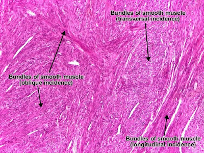
Histology and Explanation of leiomyoma Uterus

Uterine leiomyoma is a benign connective tissue tumor of the smooth muscle cells of the myometrium. Tumor cells resemble normal cells (uniform, elongated, spindle-shaped, with a cigar-shaped nucleus) and form bundles with different directions (whirled). The tumor may present areas of fibrosis, calcification and/or hemorrhage. The tumor is well circumscribed, but not encapsulated.
Weekend Case: This rash started on a 55-year-old man's forearms and legs and spread to his trunk ...
What is the Diagnosis and Treatment?


Answer:
Rocky mountain fever............The disease is caused by Rickettsia rickettsii, a species of bacterium that is spread to humans by Dermacentor ticks. Initial signs and symptoms of the disease include sudden onset of fever, headache, and muscle pain, followed by development of rash Petechial rash Abdominal pain Joint pain severe manifestations of this disease may involve the respiratory system, central nervous system, gastrointestinal system, or renal system.
Friday, March 04, 2011
Top 10 Pathology MCQs of the week
(A) air embolism from the compound fracture
(B) bone marrow embolus from the fractured femur
(C) fat embolism from the fractured femur
(D) systemic thromboemboli from the left atrium
(E) venous thromboemboli from the deep leg veins
2- A 67-year-old woman presents with a 4-week history of headaches, facial pain, blurred vision, and intense pain and stiffness in her shoulders and hips. She is diagnosed with a vasculitis, and a biopsy of an affected artery is taken. Histological examination is most likely to reveal which of the following characteristic findings?
(A) concentric “onion skin” thickening and fibrosis
(B) extensive intra- and extravascular granulomatous inflammation
(C) fragmentation of the internal elastic lamina with giant cells
(D) hyaline arteriolosclerosis and luminal narrowing
(E) segmental fibrinoid necrosis and neutrophil infiltration
3- A 67-year-old retiree was employed for many years in the plastics industry where he was exposed to vinyl chloride. This industrial exposure has increased his likelihood of developing which of the following?
(A) focal nodular hyperplasia
(B) hepatic adenoma
(C) hepatic angiosarcoma
(D) hepatic fibroma
(E) hepatocellular carcinoma
4- A 2-day-old male infant has not passed any meconium and is now developing signs of obstruction. Examination of the colon would reveal which of the following abnormalities?
(A) absence of parasympathetic ganglion cells in the submucosal and myenteric plexus
(B) absence of the nerve fibers that innervate the wall
(C) atrophy of the mucosal lining of the wall
(D) hypertrophy of the muscle coat of the wall
(E) presence of multiple small polyps along the mucosal surface
5- A 57-year-old woman had her last menstrual period at the age of 46. However, for the past 4 months she has experienced intermittent vaginal hemorrhage. A right ovarian mass is identified.Which of the following is the most likely diagnosis?
(A) arrhenoblastoma
(B) Brenner tumor
(C) dysgerminoma
(D) granulosa cell tumor
(E) Sertoli-Leydig cell tumor
(F) teratoma
6- A 28-year-old recently divorced man with no significant past medical history presents to the mergency room with progressive lower abdominal pain and cramping over the past 4 days, both of which are relieved by defecation. He has suffered from substantial bloody and mucoid diarrhea during this time. His
temperature is 102.8°F. Laboratory studies reveal an elevated white blood cell (WBC)count and a high erythrocyte sedimentation rate. Sigmoidoscopy reveals extensive rectal and sigmoid hyperemia and edema, numerous superficial ulcerations, and small focal mucosal hemorrhages, many of which have
suppurative centers. Significant intestinal narrowing is seen in the distal transverse colon.These findings most likely suggest a diagnosis of which of the following?
(A) amebic colitis
(B) collagenous colitis
(C) cytomegalovirus enterocolitis
(D) pseudomembranous colitis
(E) ulcerative colitis
7- A50-year-old woman complains of a lump in her thyroid gland. Fine-needle aspiration of
this lump is reported as medullary carcinoma. A test of her serum would most likely reveal an increased level of which of the following?
(A) alpha-Fetoprotein
(B) cancer antigen (CA) 125
(C) CA 15–3
(D) calcitonin
(E) human chorionic gonadotropin
8- A 14-year-old boy is referred to you for nasal obstruction and frequent nosebleeds. Which of
the following is the most likely diagnosis?
(A) inverted papilloma
(B) juvenile angiofibroma
(C) nasal polyps
(D) nasopharyngeal carcinoma
(E) wegener granulomatosis
9- A 52-year-old woman presents to her primary care physician complaining of increasing fatigue
and mild shortness of breath. Blood work reveals a hypochromic anemia with a hemoglobin
concentration of 10.4 g/dL, MCV of 76 μ/m3,MCHC of 29 g/dL, and a decrease in the absolute reticulocyte count. WBC and platelet counts are within normal limits. Serum iron and ferritin levels are low and total iron-binding capacity is elevated. Which of the following conditions best accounts for these findings?
(A) anemia of chronic disease
(B) aplastic anemia
(C) hypothyroidism
(D) iron deficiency
(E) pernicious anemia
10- A 52-year-old woman has had rheumatoid arthritis for many years. She now comes to you
complaining of the development in the past few months of redness, burning, and itching of
her eyes and a dry mouth, making swallowing difficult. This newly developing condition
gives the patient a greatly increased risk for which of the following?
(A) esophageal carcinoma
(B) leukemia
(C) lymphoma
(D) melanoma
(E) pleomorphic adenoma
Answers:
1.d
2.e
3.e
4.a
5.b
6.e
7.d
8.c
9.d
10.a
Important Questions of Anatomy MBBS 1st Professional (Part-1) Exam
Name the different types of bones in axial and appendicular skeletons?
Classify synovial joints? Give examples?
Describe microscopic appearance of different types of cartilage. Give their examples.
Draw and label the diagram of microscopic appearance of hairy skin. Mention its appendages.
Describe placenta and enlist its functions.
Draw and label transverse section of midbrain at the level of superior colliculus. What is the functional significance of superior colliculus?
Describe the boundaries and contents of femoral triangle?
Name the invertors and evertors of foot. Give their nerve supply.
Define supination and pronation. What are the muscles and nerves involved. At which joint these movements take place?
Describe the structural classification of bones with examples. What are the function of bones.
What are the characteristics of a synovial joint? Name the synovial joints of upper limb?
Name different components of cell. Briefly describe their functions/
What are the types of cartilage found in humans body? Draw and label the microscopic appearance of hyaline cartilage?
Define following: Capacitation, Fertilization, Zonal Reaction
What are the rotator cuff muscles? Describe their actions and nerve supply?
Describe the boundaries and contents of cubital fossa?
Name the glutei muscles? Give their actions and nerve supply?
Describe the arches of foot. Name the factors which are responsible for maintenance of these arches.
Describe briefly the coronary circulation?
Give a brief account of anatomical planes of the body?
Briefly describe cartilaginous joints?
Describe the cell cycle?
Describe the striation and functions of cilia?
Describe microscopic structure of adipose tissue and its functions?
Describe microscopic structure of neutrophils and their functions?
Describe microscopic structure of Cardiac muscle?
Name the encapsulated nerve endings and briefly describe the pacinian corpuscles?
Describe the microscopic structure of splenic pulp?
Describe oogeneisis?
Define cortical and zona reactions? What is their role in fertilization?
Discuss implantation? Name abnormal sites of implantation?
Describe the development of neural tube?
Name the derivatives of ectoderm?
What are important morphological feature of 5 weeks old embryo?
What are various parts of decidua and what is their fate?
Describe briefly placental circulation?
Name common viral infections which may cause congenital malformation. Discuss role of rubella virus as teratogen?
Define following terms : phenotype, karyotype, Alleles
Describe medial longitudinal bundle?
Name the basal ganglia and describe caudate nucleus?
What is striate cortex, give its connections?
Define cisterns, give their names and describe cisterna pontis?
Describe important features of olivary complex?
Give an account of formation, course and termination of fornix?
Name the arteries which supply superolateral surface of brain and describe anterior cerebral artery?
Name the deep nuclei of cerebellum and describe dentate nucleus?
Describe external feature of thalamus?
Give an account for lymphatic drainage of lower limb?
Give origin, insertion, actions and nerve supply of gluteus maximus muscle?
Describe femoral canal and give its clinical importance?
Describe the capsule of knee joint, name its ligaments, what are the openings in this capsule?
Give an account of course and relations of tibial nerve, name its terminal branches?
Describe deltoid ligament of ankle joint? What is its role in strengthing of this joint?
Give an account for anastomsis around knee joint?
Give an account for origin, insertion, nerve supply and actions of 1st layer of muscles of sole?
Describe extensor retinacula?
Name the movements of foot? Give a brief account of joints at which inversion and eversion take place?
Stress Management Tips for Medical Students
When you're feeling job and school related stress, it's easy to feel like you're alone. It can be especially frustrating to see classmates who seem to whiz through their studies without being fazed by stress. If you're feeling like you're the only student in your medical school class who is feeling overwhelmed, there's a very good chance that you are wrong.
2- Physician Heal Thyself:
It isn't going to do you or your future patients any good if you neglect your own well being. You need to remind yourself regularly that a healthy body is much better equipped to deal with a medical career than one that is unhealthy.
3- Avoid Med Student Hypochondria:
you do need to stay in touch with your own physical condition, you can't diagnose yourself with every disease that you study.
4-Get Mentally Prepared for the Job:
It's important to prepare yourself for what dealing with real patients is going to be like. The stress of dealing with sickness and death is very real, and it's a necessary part of working in the medical profession,You'll be much better equipped for the reality of working in a hospital if you prepare yourself by mastering stress.
Thursday, March 03, 2011
Medical Blunder: Diagnosis of Alcohol Consumption

SYMPTOM: Drinking fails to give taste and satisfaction, beer is unusually pale and clear.
FAULT: Glass empty.
ACTION: Find someone who will buy you another beer.
SYMPTOM: Drinking fails to give taste and satisfaction,and the front of your shirt is wet.
FAULT: Mouth not open when drinking or glass applied to wrong part of face.
ACTION: Buy another beer and practice in front of mirror. Drink as many as needed to perfect drinking technique.
SYMPTOM: Feet cold and wet.
FAULT: Glass being held at incorrect angle.
ACTION: Turn glass other way up so that open end points toward ceiling.
SYMPTOM: Feet warm and wet.
FAULT: Improper bladder control.
ACTION: Go stand next to nearest dog. After a while complain to the owner about its lack of house training and demand a beer as compensation.
SYMPTOM: Floor blurred.
FAULT: You are looking through bottom of empty glass.
ACTION: Find someone who will buy you another beer.
SYMPTOM: Floor moving.
FAULT: You are being carried out.
ACTION: Find out if you are being taken to another bar. If not, complain loudly that you are being kidnapped.
SYMPTOM: Opposite wall covered with ceiling tiles and fluorescent lights across it.
FAULT: You have fallen over backward.
ACTION: If your glass is full and no one is standing on your drinking arm, stay put. If not, get someone to help you get up.
SYMPTOM: Everything has gone dim, mouth full of cigarette butts.
FAULT: You have fallen forward.
ACTION: See above.
SYMPTOM: Everything has gone dark.
FAULT: The Bar is closing.
ACTION: Panic.
Download Orthopedics Lecture Notes Power Point Presentations
1 - General Complications of Fractures -
2 - Local Complications of Fractures -
3 - Late Complications of Fractures -
4 - Late Soft Tissue Complications -
5 - Hand Part1 -
6 - Hand Part2 -
6 - Hand Deformities, Fractures, and Palsy -
7 - Knee Joint -
9 - Ankle & Foot
10 - Osteomyelitis
11 - Septic Arthritis
12 - Bone Tumors -
13 - Bone Cancer
15 - Hip Joint
16 - Hip Joint Disorders
17 - Developmental Dysplasia of The Hip
18 - Anatomy of The Spine
19 - The Spine - Hx & PE
20 - Scoliosis & Kyphosis
21 - Spondylolisthesis
22 - Shoulder Joint -
23 - Shoulder Dislocation
24 - The Elbow
25 - Principles of Fracture & Treatment
Study skills for medical school students

Hurdling medical school is a massive task. It does not only require intellect, perseverance and hard work, but also the correct study skills. You have to adapt a smart strategy to maximize your learning process. Here are some study skills that you can use:
1-Do not cram: Cramming is never advisable. This is the major cause of mental block. If you really want to learn, then you have to study everyday. Develop a genuine interest in your subjects and you'll be motivated to learn more.
2- Understand what you are memorizing: Read the subject matter and then summarize in your own words. You should be able to explain using your own terminology.
3- Make use of mnemonics: Get the first letters of the data to be memorized and form a memorable word that you could easily remember. i.e.FOG for F-flow of blood, O- oxyhemoglobin, G - globin.
4- Keep your notes well organized: Make use of highlighters. Highlight the major points with a red pen and then the minor points with perhaps, a pink color. Rewrite your notes into a neater and cleaner page.
5- Mentoring by your professor or others: If you still could not cope up with the lessons, then you can ask help from your classmates or from your professor himself. You could also initiate a group study where you meet with your classmates and discuss one topic per session.
Wednesday, March 02, 2011
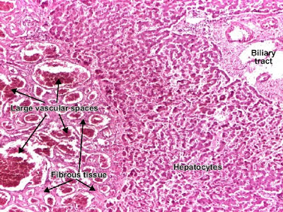
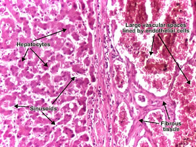
Histology and explanation of Cavernous hemangioma liver

Cavernous hemangioma is a benign connective tissue tumor resulted from endothelial cells proliferation. It is a non-encapsulated tumor, with an infiltrative, lobular growing. The tumor consists in large (cavernous) spaces, lined by tumor endothelial cells (which appear very similar to normal cells). These interconnected spaces are filled with blood and separated by a fibrous tissue.


