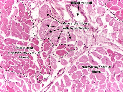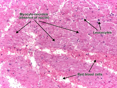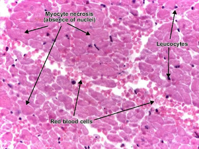
Recent myocardial infarct (in the first 12 - 24 hours): myocardial fibers are still well delineated, with intense eosinophilic (pink) cytoplasm, but lost their transversal striations and the nucleus. The interstitial space may be infiltrated with red blood cells. Make the distinction between interstitial leucocytes (small, outside the myocardial fibers) and the myocardial nucleus (should be central and unique, but is absent here).

Recent myocardial infarct (first 24 hours) (detail).


0 comments:
Post a Comment