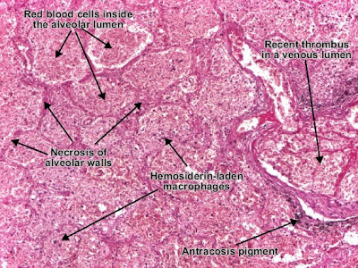
Pulmonary infarct (hemorrhagic infarct of the lung) is an area of ischemic necrosis produced by venous thrombosis on a background of passive congestion of lung. In infarct area, alveolar walls, vascular walls and bronchioles are necrotic. They appear eosinophilic (pink), homogenous, lacking the nuclei, but keep their shapes - "structured necrosis". Alveolar lumens from infarcted area are invaded by red blood cells - hemorrhagic infarct (red).

Pulmonary infarct in an area of passive congestion. The hemosiderin-laden macrophages present inside the alveolar lumen are witnesses of pre-existent passive congestion.

Pulmonary (hemorrhagic) infarct produced by venous thrombosis.


0 comments:
Post a Comment